Home

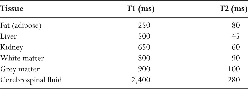
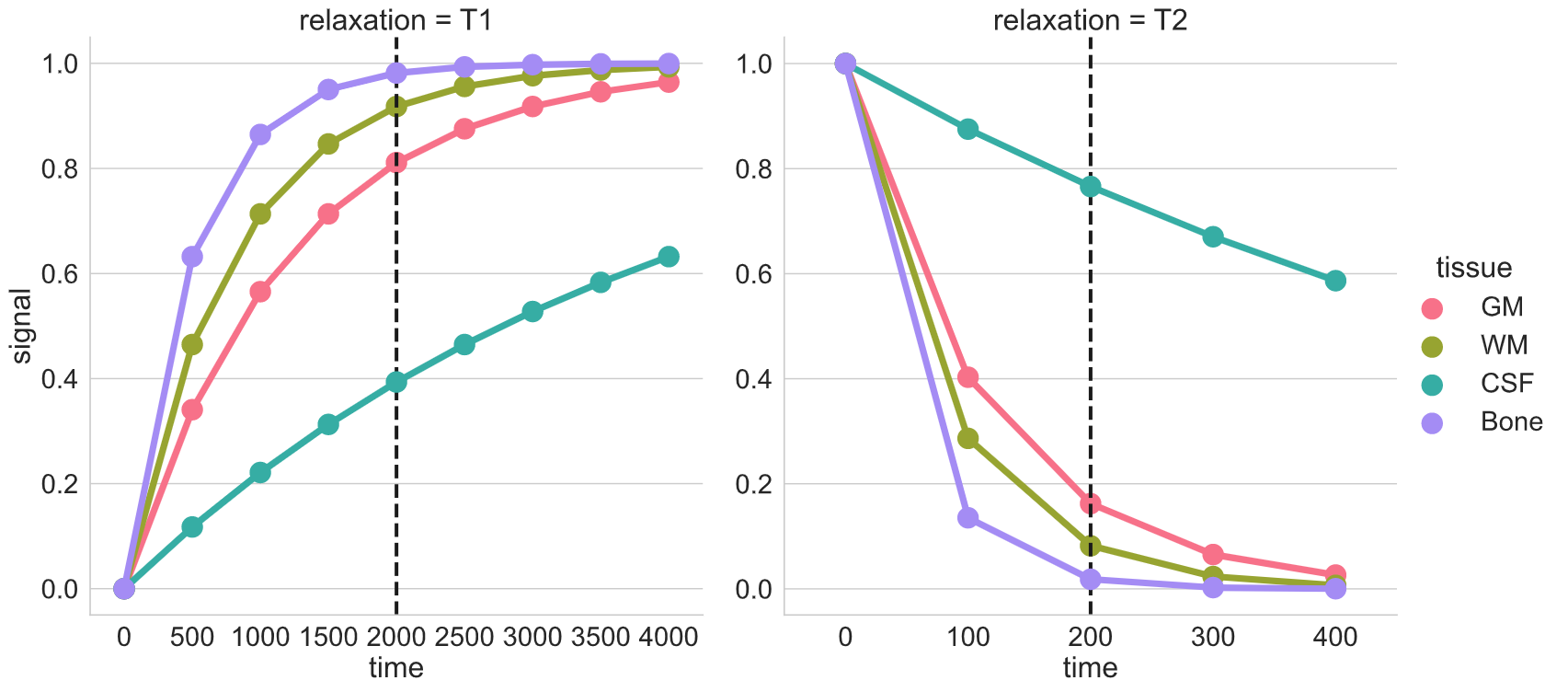
switch Represent Mission t1 and t2 values for different tissues violinist episode Archaeologist

T1 and T2 relaxation times at various anatomical regions at WM and GM... |  Download Table
T1 and T2 relaxation times at various anatomical regions at WM and GM... | Download Table

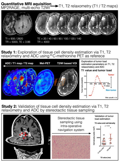
Cancers | Free Full-Text | Magnetic Resonance Relaxometry for Tumor Cell  Density Imaging for Glioma: An Exploratory Study via 11C-Methionine PET and  Its Validation via Stereotactic Tissue Sampling
Cancers | Free Full-Text | Magnetic Resonance Relaxometry for Tumor Cell Density Imaging for Glioma: An Exploratory Study via 11C-Methionine PET and Its Validation via Stereotactic Tissue Sampling

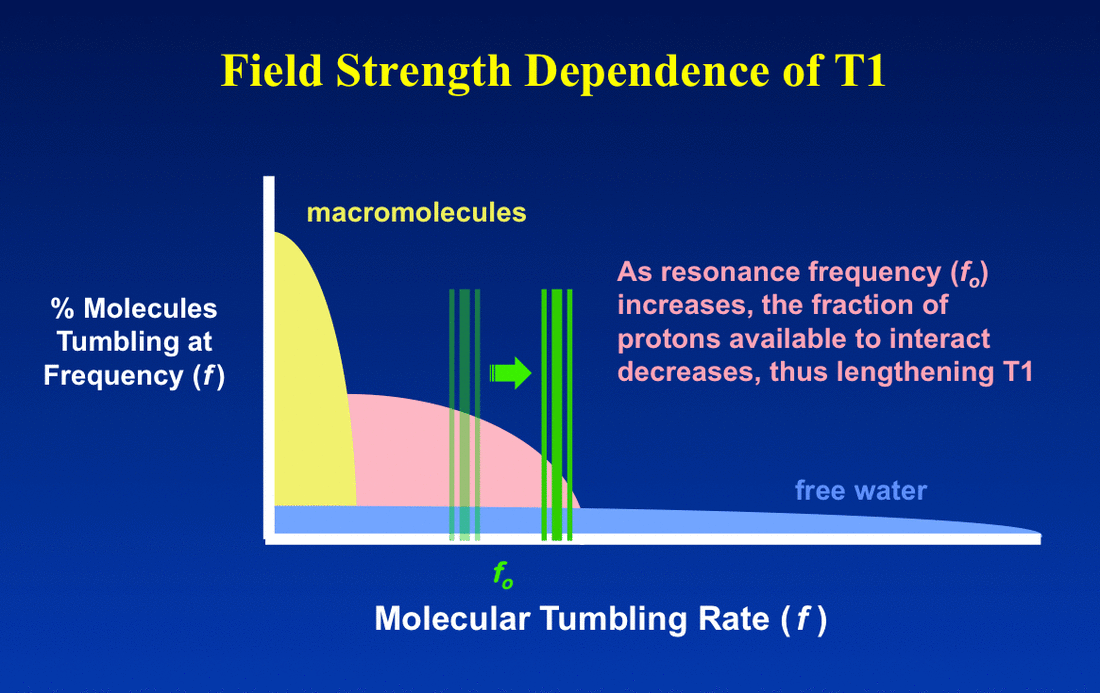
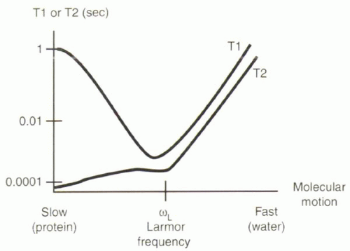
Field strength dependence of T1 & T2 - Questions and Answers ​in MRI

Magnetic Resonance | Radiology Key

T1 and T2 Values 0.5T Tissues FAT LIVER WHITE M GRAY M CSF ppt download

Breast Tissue T1 and T2 Values at 1.5 Tesla | Download Table

Magnetic Resonance Imaging

What are normal relaxation times of tissues at 3 T? | Semantic Scholar

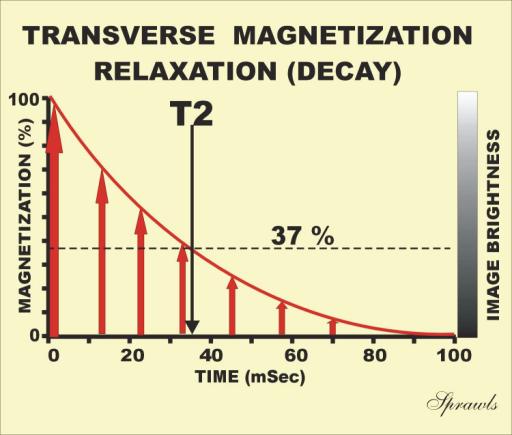
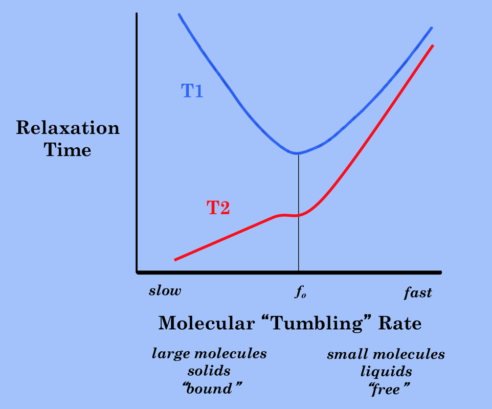
Relaxation time, T1, T2 - Questions and Answers ​in MRI

3: Some approximate T1, T2, T2 * and proton concentration values for... |  Download Scientific Diagram
3: Some approximate T1, T2, T2 * and proton concentration values for... | Download Scientific Diagram

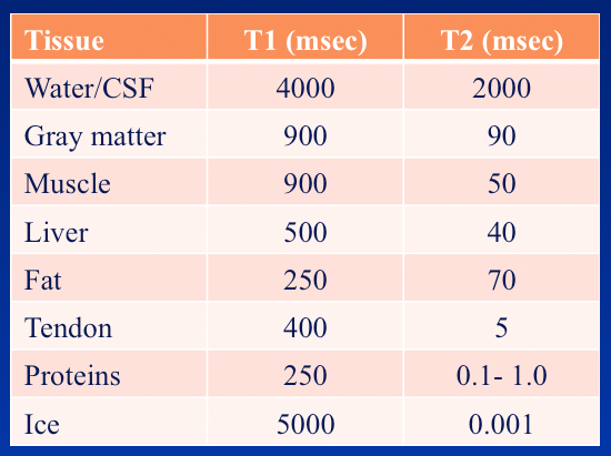
Typical T1 and T2 values for various body tissues* | Download Table

Overview of Structural MRI (Pre)processing and Neuroimaging Analysis

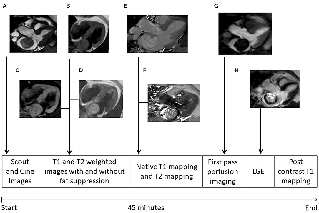
Frontiers | Cardiac Myxomas Show Elevated Native T1, T2 Relaxation Time and  ECV on Parametric CMR
Frontiers | Cardiac Myxomas Show Elevated Native T1, T2 Relaxation Time and ECV on Parametric CMR

PDF] MR imaging: possibility of tissue characterization of brain tumors  using T1 and T2 values. | Semantic Scholar
PDF] MR imaging: possibility of tissue characterization of brain tumors using T1 and T2 values. | Semantic Scholar
T1 and T2 values for the training tissue. | Download Table

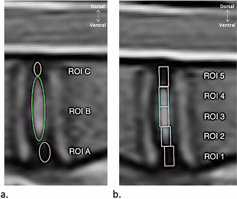
Comparison of MRI T1, T2, and T2* mapping with histology for assessment of  intervertebral disc degeneration in an ovine model | Scientific Reports
Comparison of MRI T1, T2, and T2* mapping with histology for assessment of intervertebral disc degeneration in an ovine model | Scientific Reports

T1 and T2 values at 1T. | Download Table

Relaxation time, T1, T2 - Questions and Answers ​in MRI

Typical T1 and T2 values for various body tissues* | Download Table

Table 1 from Normal tissue quantitative T1 and T2* MRI relaxation time  responses to hypercapnic and hyperoxic gases. | Semantic Scholar
Table 1 from Normal tissue quantitative T1 and T2* MRI relaxation time responses to hypercapnic and hyperoxic gases. | Semantic Scholar

T1 and T2 signal - Radiology Cafe

Tissue Characteristics and Contrast Agents: PD, T1, and T2 | Thoracic Key